The direct interface between the human brain and the computer promises many interesting perspectives, but, above all, it will help people with disabilities. Returning movement to paralyzed limbs, helping to stand up and working with a computer – all this today is no longer a fantasy, although it is often associated with surgical intervention in the living brain in the form of electrode implantation. But there is a good replacement for this without serious surgery – ultrasound.
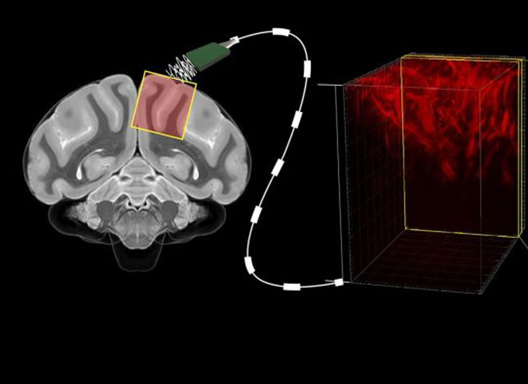
A combined team of American, Chinese and Russian scientists have developed and tested a technology for recognizing brain activity using functional ultrasound technology in primates. With minimal surgical intervention – it was only required to remove a small area in the cranium – it was possible to recognize the activity of neurons with a resolution of 100 microns. This is an order of magnitude larger than the size of one neuron (about 10 microns), but without the introduction of electrodes into the brain tissue.
In fact, scientists with minimal surgical intervention were able to obtain a map of brain activity in real time without bulky equipment in the form of MRI scanners and with a resolution close to the capabilities of electrodes embedded in the brain. This was the first point of the completed program.
In the second stage, the researchers were able to link the muscular activity of primates to the brain activity map and learned to literally read thoughts – to predict the movements of animals before they began to act. This required loading into the neural network data on brain activity, taken using electrodes in past long-term studies, and correlating with it the muscle response (activity) of the experimental primates. After that, the trained neural network was provided with data on brain activity captured by ultrasonic sensors, and the algorithm was able to predict the movements of animals before they made it.
We add that a map of brain activity using functional ultrasound is created by fixing the power of blood flow in the vessels of the brain. Ultrasound sensors respond to red blood cells, the movement of which creates high-frequency reflections. When blood flow increases in the observed area, this means that a group of neurons begins to assign a task to certain muscles. The AI instantly interprets brain activity into muscle activity and can, for example, force the prosthesis to do what the subject wishes.
Based on the results obtained, studies were started with the involvement of volunteers from among people with open craniocerebral injuries. In such cases, the skull does not need to be specially removed. There is already a “window” in the head for the operation of ultrasonic sensors. In the future, to organize such an interface, it may be sufficient to simply implant ultrasonic transducers under the cranial bone. Certainly, this does not compare with the burial of electrodes in the brain.
If you notice an error, select it with the mouse and press CTRL + ENTER.


